4 Contrast: TR, TE, and T1/T2 Weighting
4.1 In a nutshell
In MRI, choosing when to repeat (TR) and when to listen (TE) determines whether you see differences in T1 or differences in T2.
| Term | Meaning |
|---|---|
| TR (repetition time) | The time from one RF pulse to the next RF pulse |
| TE (echo time) | The time from the RF pulse until the signal (echo) is received |
| PD (proton density) | The amount of hydrogen at that location |
4.2 The signal equation (spin echo, simplified)
\[ S = PD \times \underbrace{\left(1 - e^{-TR/T_1}\right)}_{\text{how much longitudinal magnetization recovered by TR}} \times \underbrace{e^{-TE/T_2}}_{\text{how much transverse magnetization remains at TE}} \]
- Short TR → longitudinal recovery is incomplete → only tissues with short T1 (fat) give large signal → T1-weighted
- Long TE → after the transverse signal has decayed → only tissues with long T2 (water) keep their signal → T2-weighted
- Long TR and short TE → neither T1 nor T2 has an effect → the amount of hydrogen itself → PD-weighted
| TR | TE | Appears bright | Appears dark | |
|---|---|---|---|---|
| T1-weighted | Short (~500 ms) | Short (~10 ms) | Fat | Water |
| T2-weighted | Long (~4000 ms) | Long (~100 ms) | Water, edema | Muscle |
| PD-weighted | Long | Short | Hydrogen-rich areas | Bone (cortex) |
4.3 Demo: move TR and TE
Schematic on the left: outer ring = subcutaneous fat, middle = muscle, upper-left circle = water, lower right = bone (with fatty bone marrow inside). Brightness is displayed with the strongest signal among the three tissues shown as white.
Let’s try:
- TR = 500, TE = 10: fat and bone marrow are bright (T1-weighted)
- TR = 4000, TE = 100: water is bright and muscle is dark (T2-weighted)
- TR = 4000, TE = 10: the differences become small (PD-weighted)
4.4 Python: make three kinds of weighted images
code/04_contrast.py
# Use the spin-echo signal equation to make T1-, T2-, and PD-weighted images of the same phantom
import numpy as np
import matplotlib.pyplot as plt

N = 200
y, x = np.mgrid[-1:1:N * 1j, -1:1:N * 1j]


def disk(cx, cy, r):
    return (x - cx) ** 2 + (y - cy) ** 2 < r ** 2


# tissue labels: 0=background, 1=fat, 2=muscle, 3=water, 4=cortical bone
label = np.zeros((N, N), int)
label[disk(0, 0, 0.9)] = 1
label[disk(0, 0, 0.78)] = 2
label[disk(-0.3, -0.25, 0.18)] = 3
label[disk(0.35, 0.15, 0.15)] = 4
label[disk(0.35, 0.15, 0.09)] = 1  # bone marrow (fat)

#            PD    T1[ms]  T2[ms]
props = {1: (0.90, 400, 100),
         2: (0.75, 1400, 45),
         3: (1.00, 4000, 2000),
         4: (0.05, 1000, 1)}


def spin_echo(TR, TE):
    S = np.zeros((N, N))
    for lab, (pd, T1, T2) in props.items():
        S[label == lab] = pd * (1 - np.exp(-TR / T1)) * np.exp(-TE / T2)
    return S


settings = {"T1w (TR=500, TE=10)": (500, 10),
            "T2w (TR=4000, TE=100)": (4000, 100),
            "PDw (TR=4000, TE=10)": (4000, 10)}

fig, axes = plt.subplots(1, 3, figsize=(10.5, 3.8))
for ax, (title, (TR, TE)) in zip(axes, settings.items()):
    ax.imshow(spin_echo(TR, TE), cmap="gray")
    ax.set_title(title)
    ax.axis("off")
fig.tight_layout()
fig.savefig("figs/04_contrast.png", dpi=150)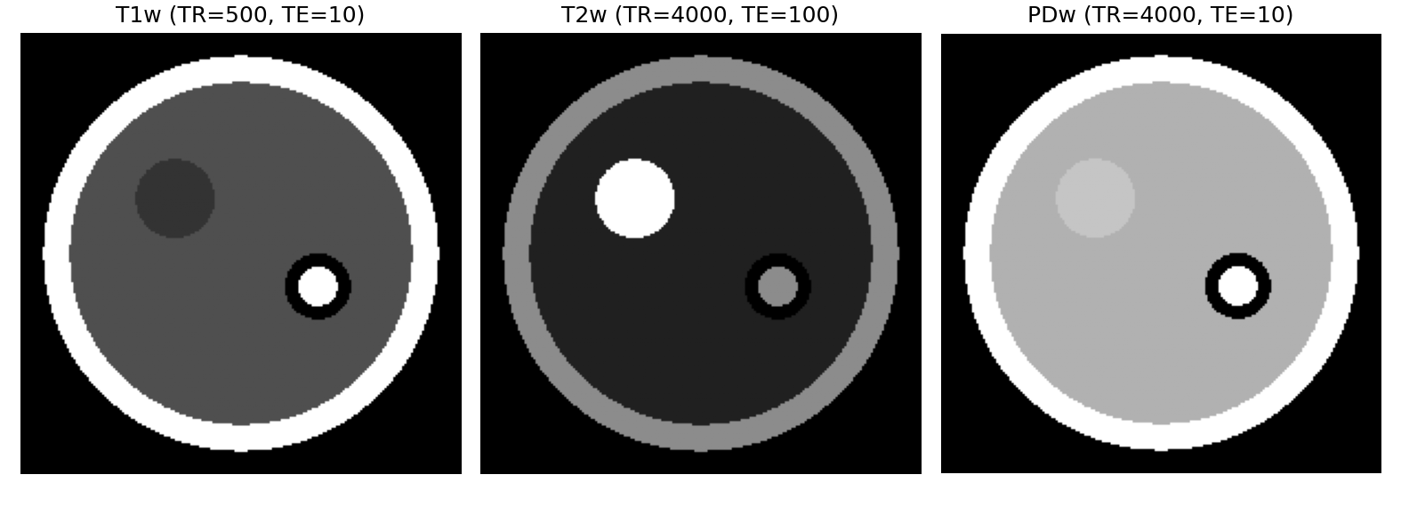
4.5 Quantitative MRI: measuring “values,” not “weighting”
The brightness of T1- and T2-weighted images is a relative value that changes with the scanner and settings. In research, it is common to acquire many images with different TE or TI and create images in which the T1 or T2 value (ms) is estimated for each pixel (T1 maps, T2 maps).
- T2 map: acquire many echoes at different TEs (multi-echo) and fit \(S(TE) = S_0 e^{-TE/T_2}\) pixel by pixel (implemented in Chapter 8)
- T1 map: estimated from inversion recovery (IR) with different TIs, or from acquisitions with different flip angles
- MR fingerprinting (MRF): acquire while varying the conditions pseudo-randomly, then match against a dictionary to estimate T1, T2, fat fraction, and so on all at once (Ma et al. 2013). It has also been used to measure muscle water T1 and fat fraction after exercise (Höger et al. 2023)
4.6 Summary
- TR is the knob that brings out T1 differences; TE brings out T2 differences
- Short TR, short TE = T1-weighted (fat is bright); long TR, long TE = T2-weighted (water is bright)
- In research, people often measure the T1 and T2 values themselves (maps) rather than weighted images